Looking for a needle in a haystack meaning of a phraseological unit. Separation based on charge, size and shape of molecules
Like a needle in a haystack. Express So it's impossible to find. The partisans, like a needle in a haystack, were lost in the forest, but every night they made themselves felt, and very sensitively(N. Biryukov. Seagull).
Phraseological dictionary of the Russian literary language. - M.: Astrel, AST. A. I. Fedorov. 2008.
See what “Like a needle in a haystack” is in other dictionaries:
like a needle in a haystack- Like (as if, as if) a needle in a haystack (disappear, get lost) About whom, what, who (what) cannot be found... Dictionary of many expressions
needle- And; pl. genus. lok, dat. lkam; and. = needle 1), 2), 3), 4) Sew silk with a thin needle. Needle/bow for a sewing machine. A needle from a gramophone. Prickly pine needles. Soft cactus needles... Dictionary of many expressions
needle- And; pl. genus. lok, dat. lkam; and. = Needle (1 4 digits). Sew silk with a thin needle. I. for a sewing machine. I. from the gramophone. Prickly pine needles. Soft cactus needles. Hedgehog needles. * Where the needle goes, so does the thread (Last.). ◊ From a needle. = Brand new. Not a needle who... Encyclopedic Dictionary
stack- A; sentence, about a hundred/ge, in a stack/ and in a hundred/ge; pl. haystacks/; m. see also. haystack, stack A large pile of hay, straw or unthreshed bread, round or quadrangular in shape, tapering towards the top and folded in the open air for storage. Haystack... Dictionary of many expressions
be invisible- ▲ to stand out (than) little invisibility. inconspicuous, slightly prominent. unnoticed. inconspicuous. inconspicuous. imperceptible. elusive (# aroma). insensible. insensitive. intangible (# results). invisible hide (#… … Ideographic Dictionary of the Russian Language
stack- A; sentence about a haystack, in a haystack and in a haystack; pl. haystacks; m. A large pile of hay, straw or unthreshed bread, round or rectangular in shape, tapering towards the top and piled in the open air for storage. C. hay, straw. Carry haystacks to the haystack... Encyclopedic Dictionary
needle- and, gen. pl. lok, dat. lkam, w. Same as needle (in 1, 2, 3 and 4 digits). Where the needle goes, so goes the thread. Saying. [Anyuta] silently wrapped her embroidery in paper and collected the threads and needles. Chekhov, Anyuta. Samorodov busily opened the gramophone, wound up the spring and... ... Small academic dictionary
stack- ah, sentence about a haystack, in a haystack and in a haystack, pl. haystack, m. A large pile of hay, straw or unthreshed bread, piled in the open air for storage. Saddled horses stood in the bushes, plucking fragrant hay from a fresh stack. Sholokhov Sinyavsky,... ... Small academic dictionary
MUCH - LITTLE- Rarely, but accurately. Once, yes much. There is a parable shorter than a bird's nose (and a good one). And one eye, but a keen one, you don’t need forty. And one cow, and it’s healthy to eat. The river is shallow, but the banks are steep. The flow is not wide, but it holds. Not big, but the caftan is wide and short.... ... V.I. Dahl. Proverbs of the Russian people
In general, needles in a dream mean troubles or things that you don’t feel like doing. A dull needle, both in life and in a dream, cannot do much harm, but it does not do any good. This dream suggests that a loved one will soon become indifferent to you.
Pulling a needle out of some part of the body in a dream means that obstacles in business are causing you a lot of trouble and problems, but after such a dream everything should change - you will feel relief.
Buying needles in a dream means reconciliation with a friend. A needle and thread in a dream means that your relationship with a loved one or partner will be like a thread and a needle. Where the needle goes, so goes the thread.
The thread always follows the needle. Try to figure out who is meant by the thread and who is meant by the needle. Such a dream can also predict that you will try to achieve the same success as another person. The length of the thread in this case indicates how close your relationship with the other person will be. See interpretation: threads.
If you dream that you pricked yourself with a needle, then expect a quarrel with your loved ones. See interpretation: prick.
A dream in which you saw that you had lost a needle means the loss of a friend or loved one. Looking for a needle means wasted effort. It’s not for nothing that there is a saying “looking for a needle in a haystack.”
Finding a needle in a dream is an indication of the danger that threatens you, which will come from where you do not expect it. Searching and finding a needle is a good dream. It means that you will soon find new friends.
A broken needle in a dream means a break in a relationship with a loved one. After such a dream, expect great experiences and loneliness.
A dream in which you saw yourself working with a needle means: expect a quarrel with a loved one. For spouses, such a dream predicts that their family life will soon crack.
Interpretation of dreams from the Family Dream BookThe V. Potanin Charitable Foundation has a large program of support for young university teachers who successfully combine teaching and scientific work, which has been running for several years. A special competition has been organized to distribute grants. There are many requirements for applicants, but among other things, young teachers must give a popular science lecture on their specialty to senior students. The implementation of this wonderful idea, on the one hand, makes it possible to understand whether the applicant knows his subject, on the other hand, there are quite obvious benefits from such lectures: students broaden their horizons, receiving information on related and sometimes completely distant specialties. The Foundation gave the editors of Science and Life the opportunity to get acquainted with all the lectures given by grant applicants. We have published some of them. We were attracted to these lectures by the simplicity and clarity of presentation (lecture “A smile will make everyone brighter”, “Science and Life” No. 3, 2009), the importance of the topic (“When leaving, turn off the light!”, “Science and Life” No. 6 , 2009), a modern idea of long-known things (“Hope and support”, “Science and Life” No. 8, 2009), an unexpected look at seemingly obvious phenomena (“Biological signal fields...” , “Science and Life” No. 1, 2009). We present to our readers a lecture given by Lyudmila Trukhacheva, Candidate of Pharmaceutical Sciences, Associate Professor at the Moscow Medical Academy. I. M. Sechenov. The story is truly detective...
On an early August morning in 1961, hundreds of crazed birds attacked the seaside town of Capitola in the US state of California. Hitherto harmless gray petrels, in flocks and individually, crashed into windows and walls of houses at high speed, dived into street lamps and attacked passers-by. This incident inspired Alfred Hitchcock's film The Birds.
A quarter of a century later, in the winter of 1987, another mysterious story happened on Prince Edward Island off the North Atlantic coast of Canada: more than a hundred people became victims of severe food poisoning. It turned out that all the victims ate blue mussels. In addition to the usual symptoms - vomiting, cramps, diarrhea and headache - patients experienced disorientation, a feeling of panic, amnesia, and in some cases, seizures and coma. Almost all of them showed symptoms of a mental disorder; patients showed uncontrolled aggressiveness, often accompanied by crying or laughter. Unfortunately, three victims could not be helped - they died in the first days. More than a quarter of the other victims had impaired short-term memory. They could not remember anything that happened after the poisoning, some did not recognize their loved ones.
It later turned out that both cases - the first with “crazy birds” and the second with “poisoned mussels” - were the result of exposure to the same toxic agent. The condition it causes is now known as amnestic shellfish poisoning syndrome (ASP). However, there have been no previous reports of mussel food poisoning with such neurological consequences.
To clarify all the circumstances of what happened, as well as to prevent similar cases, the Canadian Department of Fisheries commissioned a group of marine biologists and chemists to isolate and identify the toxic agent.
Initial testing of the mussels for known bacterial and viral pathogens was inconclusive. Tests for heavy metals and pesticides also came back negative. The samples taken for analysis included thousands of different chemical compounds. How can one isolate one component from such a complex mixture without knowing anything about its physical or chemical properties? The task is no easier than finding a needle in a haystack.
Let's assume that we have the ability to determine whether there is a needle in the stack or not. Then the search algorithm will be as follows. First, we divide the stack into two halves and check whether there is a needle in one of the parts. If not, discard this half, divide the remaining half in half and look for the needle in the next halves. Such “split-drop-divide” manipulations will ultimately lead to the fact that the last remaining part will be nothing more than the desired needle. The main strategy of the researchers, who were faced with the task of finding and isolating the toxin, was built according to the same scheme.
First of all, it was necessary to develop a test that would reliably indicate the toxicity of the objects being studied. And here there were experiments on animals. It was found that mice exhibit the most characteristic reaction to the toxin. After introducing small amounts of the test sample, if a toxin was present in it, the experimental animals experienced a clear neurological reaction - the mice began to uncontrollably scratch and comb their shoulders with their hind legs. The test is cruel, but in light of the tragedy that occurred, scientists had no other choice.
To separate the complex components in the poisoned mussel tissue samples, the scientists used standard physicochemical methods. Both toxic and non-toxic mussel samples were processed. This approach is necessary for subsequent comparative analysis, because any difference between the samples could provide a clue to solving the mystery.
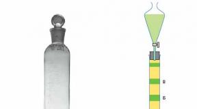
Let's follow all the steps of the process shown in the diagram and try to understand what happened at each stage.
Separation based on solubility and volatility
In the first three stages, the researchers used extraction and evaporation as a general strategy.
Extraction is the separation of a mixture of substances based on differences in solubility. Housewives are well aware of the fact that the solubility of substances in different solvents is different from the example of vanillin, which is poorly soluble in water and highly soluble in alcohol. In liquid-liquid extraction, the solute is distributed between two liquid immiscible phases. Typically one phase is water and the other is an organic solvent.

During evaporation, the solution is concentrated as a result of the evaporation of the solvent. The extract can be evaporated to a small volume and thereby increase the concentration of the analyzed component.
Now that we know the benefits of extraction and evaporation, let’s return to the search for the toxin.

To prevent possible destruction of the desired compound as a result of heating or interaction with a solvent, extraction was carried out at room temperature with an aqueous solution of methanol, a medium-strength solvent. The extraction was insufficient, but nevertheless successful: mice showed the same neurological response to the methanol extract as to the original oyster samples. The extract was then concentrated by evaporation. The separated and condensed steam was non-toxic, but the resulting residue gave the necessary reaction in mice. It became clear that poison is a non-volatile substance.
A second extraction was carried out. This time the concentrated extract was shaken with a mixture of polar and non-polar solvents. We used dichloromethane and water: these solvents do not mix and form two separate layers.
Colored substances were found in the dichloromethane fraction—phytoplankton pigments (in other words, algae). And this could already be the key to clarifying the nature of the toxin. However, the pigments themselves are not poisonous, and the dichloromethane fraction gave a negative result in experimental mice. But the toxin was present in the water layer. This suggested that the object being sought was apparently a polar, ionizable substance. Now the researchers could concentrate on the water fraction.
At the next stages, chromatographic methods of analysis were used. Here we have to go a little deeper into the theory...
Separation in motion
Chromatographic analysis, one of the most sensitive methods, first proposed by the Russian scientist Mikhail Semenovich Tsvet at the beginning of the 20th century, by the beginning of the 21st century had turned into a powerful tool, without which it is difficult to imagine analytical chemistry, and not only that.
The first experiment in separating and analyzing a substance of complex composition, carried out by M. S. Tsvet in 1903, is surprisingly simple. The researcher passed a chlorophyll solution through a tube (or, as they now say, a column) containing chalk powder, gradually diluting it with benzene. After some time, rings colored by chlorophyll components in different colors became visible in the chalk column. Having cut the column, M. S. Tsvet isolated them in their pure form and carried out a chemical analysis of each individual component.

All of us have probably been involved in chromatography in one way or another, and our parents were especially lucky in this sense. After all, in previous years, schoolchildren wrote in ink. And if a blotter fell on an ink stain, then the ink solution was divided into several “fronts” on it.
Chromatography itself is based on the distribution of one of several substances between two phases (for example, between a solid and a gas, between two liquids, etc.), with one of the phases constantly moving. The better a particular substance is sorbed (absorbed) or dissolved in the stationary phase, the lower the speed of its movement, and vice versa, the less the compound is sorbed, the greater the speed of movement. As a result, if at first we have a mixture of compounds, then gradually all of them, pushed by the mobile phase, move towards the “finish line” at different speeds and eventually separate.

After separation, all components must be identified and quantified. This is done using detectors that have little to do with the chromatographic process itself and are based on the various physicochemical properties of the substances being studied.
In modern chromatographs, the length of columns in which substances are separated reaches hundreds of meters. A few milligrams (10 -3 g) of the mixture are sufficient for analysis, and components weighing up to several picograms (10 -12 g) can be detected in it.
These are, in general terms, the basics of chromatographic analysis. Now it’s time to return to the search for a toxic agent in mussel samples.
Separation based on differences in polarity
So, column chromatography was used to separate the mixture in the remaining aqueous layer into its simple components. The sample was passed through a narrow tube containing XAD-2 resin microspheres. These microspheres retain non-polar, uncharged molecules and allow charged ions to pass through. XAD-2 is particularly effective for the separation of organic bases and acids.
Ionized acids pass through the column and exit before other organic compounds.
Of the many fractions that passed through XAD-2, only one turned out to be toxic. At the final stage, this fraction was separated using high performance liquid chromatography (HPLC). Here again, the polar solution containing the sample was passed through a column with a non-polar sorbent as the stationary phase. The resulting highly purified fraction contained all the poison of the poisoned mussels. So finally the toxin was isolated.
Separation based on charge, size and shape of molecules
However, the researchers had to make sure that the final fraction isolated by HPLC actually contained the same toxic component. To do this, it was decided to separate the aqueous fraction of XAD-2 again, but using high-voltage electrophoresis on paper.
Electrophoresis is a method of separating ions based on differences in their relative charges (the ratio of charge to mass). The ions located between the positive and negative electrodes, under the influence of the electric field, begin to move towards the electrode with a charge of opposite sign. Typically, the higher the charge-to-mass ratio of an ion, the faster it moves toward the electrode. Small, highly charged ions move ahead of large, low-charge ions. The shape of the molecules also affects the speed of movement. Thus, molecules with more concentrated charges move faster.
The researchers placed the sample on a strip of filter paper. Both ends of the strip were immersed in buffer solutions, each of which contained an electrode. During the analysis, the ions of the substances contained in the analyzed sample moved at different speeds and were separated in the form of separate stripes on the paper. In order for these stripes to “appear,” the paper was sprayed with a special reagent.
During electrophoresis of the XAD-2 fraction, an unknown band was detected next to the glutamic acid band (one of the reference samples). This band was absent in control tissue samples of nontoxic mussels. In addition, the color of the unknown band was completely different from the color of the glutamic acid band. The band containing the unknown substance was removed from the paper, and the resulting sample was injected into the HPLC column. It turned out that this sample was identical in retention time to the main substance in the final fraction separated by HPLC. In addition, this sample exhibited the same toxicity as the sample studied by HPLC.
Toxin identification by charge and mass
At the final stage, the chemical formula and molecular weight of the isolated toxin had to be determined. The problem was solved using mass spectrometry.
This method allows you to determine the composition of the molecule of a substance by measuring the ratio of the mass of ions to their charge. First, neutral molecules and atoms are converted into charged particles - ions, and then separated using the laws of motion of charged particles in a magnetic or electric field.

So, using mass spectrometry, scientists found the molecular weight
(312 g/mol) and the molecular formula (C 15 H 21 NO 6) of the isolated toxin. Spectroscopic analysis revealed the presence of double bonds and spectra characteristic of an amino group. And when comparing the spectra of the substance with spectra in an international database, the compound was identified as domoic acid.
Domoic acid is a kind of “Trojan horse” in the world of molecules. Nerve cells (neurons) mistake it for glutamic acid molecules, and this mistake becomes fatal for them. Glutamate (the ionized form of glutamic acid) is a neurotransmitter, a molecule whose responsibilities include transmitting nerve impulses from one cell to another. When a glutamate molecule binds to a glutamate receptor on the surface of a cell, the receptor opens a special channel for calcium ions to enter the cell. The influx of charges results in an electrical potential that propagates along the cell membrane and transmits excitation information to other neurons. Frequent stimulation of this mechanism can lead to the emergence of new connections between neurons, so glutamate plays a key role in the processes of thinking, learning, and memorizing information.

But an excess amount of glutamate leads to uncontrolled excitation of the cell and ultimately to its death. Moreover, this process is cascading, since overexcitation of a dying cell is transmitted along the chain to nearby neurons. Ultimately, this biochemical cascade can cause brain damage and neurodegenerative disorders.
Domoic acid is similar to glutamic acid. However, the five-membered ring contained in its structure makes the molecule less flexible than glutamine, which causes domoic acid to bind more tightly to the glutamine receptor. And as a result, its stimulating effect is 30-100 times higher.
But the question remains - how did domoic acid get into the tissues of mussels, as well as into the anchovies that birds feed off the coast of California? Here we must remember that phytoplankton pigments were found in one of the fractions after extraction. A thorough study of domoic acid led to the discovery of its carriers - needle-shaped diatoms called Pseudo-nitzschia pungens. These algae are found in all the oceans of the world and, therefore, could become the beginning of the food chain in many regions. This is how birds that ate anchovies, which in turn ate poisonous algae, became poisoned by domoic acid.
Currently, most coastal countries carry out continuous monitoring of seafood using the HPLC method in order to promptly detect the presence of domoic acid. The measures were successful, and there have been no reports of poisonings since 1987.
The solution to this detective story would hardly have been possible without modern physical and chemical methods of analysis.
Literature:
V. S. Asatiani. Chemistry of our body. - M.: Nauka, 1969, 304 p.
F. Bayburtsky. Chromatography is a simple way to analyze complex substances// Science and Life, 1998, No. 2.